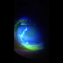
Smartphone video clip of a Herpes Simplex Virus Keratitis fluorescein pattern.
Herpes Simplex Virus Keratitis remains a clinical diagnosis based on the presence of a dendritic ulcer, as its most common presentation.
It is characterized as a “linear branching pattern with terminal bulbs, swollen epithelial borders, and central ulceration through the basement membrane”.
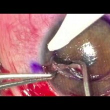
The patient is a 30 year old male with Terrien's marginal degeneration.
There was severe peripheral thining of the superior cornea, with neovascularization and lipid deposition. Best-corrected visual acuity was impaired due to irregular astigmatism.
Extensive description of the case is reported on the "Clinical Cases" section.
To prevent further progression, avoid the risk of spontaneous perforation, and to improve visual function, a surgical approach was planned and proposed to the patient.