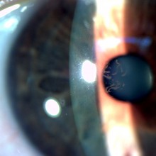
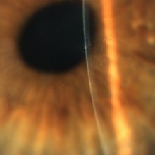
A 54-year old female patient was referred to our clinic due to long-standing progressive right eye blurred vision. No relevant personal or familial history. Right eye best-corrected visual acuity was 6/10. On biomicroscopy examination, central subepithelial spiraled opacities were characteristic of Map-dot-fingerprint Dystrophy, also known as Epithelial Basement Membrane Dystrophy or Cogan's Microcystic Epithelial Dystrophy. The disease may result in decreased vision (as in this patient) and/or recurrent corneal erosions.
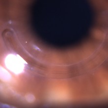
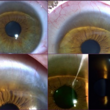
Pellucid marginal degeneration is a slowly progressive corneal ectatic condition often leading to severe visual impairment of working age people.
The peripheral corneal thinning location requires a large graft size, eventually a peripheral lamellar crescentic keratoplasty followed by a central penetrating keratoplasty.
Besides surgically more challenging, corneal transplantation is associated to increased risk of vascularization and rejection.
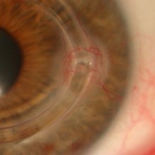
Dots, cysts and maps in the cornea of a patient complaining of suggestive recurrent corneal erosions episodes.
Named after the slit lamp appearance of corneal findings, Map-dot-fingerprint Dystrophy is the most common corneal dystrophy. Map-dot-fingerprint dystrophy, Cogan’s dystrophy, Cogan microcystic epithelial dystrophy, epithelial basement membrane dystrophy, and anterior basement membrane dystrophy all designate the same condition.
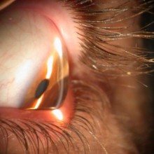
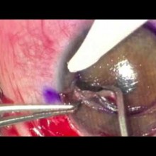
The patient is a 30 year old male with Terrien's marginal degeneration.
There was severe peripheral thining of the superior cornea, with neovascularization and lipid deposition. Best-corrected visual acuity was impaired due to irregular astigmatism.
Extensive description of the case is reported on the "Clinical Cases" section.
To prevent further progression, avoid the risk of spontaneous perforation, and to improve visual function, a surgical approach was planned and proposed to the patient.